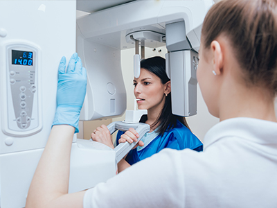
Digital radiography replaces traditional film with modern sensors and computer systems to capture detailed images of teeth, gums, and jaw structures. Instead of waiting for film to develop, clinicians can view high-resolution images almost instantly on a monitor. This immediacy helps clinicians detect issues sooner, explain findings to patients with clear visuals, and make timely decisions about next steps in care.
For patients, the experience of having dental x‑rays taken is quicker and more comfortable. Sensors are slim and designed to conform to the mouth, and the ability to preview images right away reduces the need for repeat exposures. At the same time, digital files integrate cleanly with electronic dental records, simplifying how information is stored and retrieved during future visits.
While the underlying technology is sophisticated, the practical benefit is straightforward: clearer images with a more efficient workflow that supports accurate diagnosis and a better patient experience. This technology is a foundational tool in contemporary dentistry and a standard part of preventive, restorative, and surgical planning.
Digital radiography relies on electronic sensors—either intraoral plates or extraoral detectors—that convert x‑ray energy into an electronic signal. When the sensor is exposed, it captures a pattern of varying intensities that correspond to anatomical structures. That raw data is rapidly processed by software to create a sharp, detailed image that can be adjusted for contrast, brightness, and magnification.
These images are produced without film chemistry; instead, they transfer a digital file directly to a computer. Because the data is digital, clinicians can enhance specific areas to better visualize fine details like root structure, bone levels, or early decay. That ability to manipulate images without additional radiation exposure improves diagnostic confidence and helps tailor treatment planning to each patient’s situation.
Different sensor types and imaging modes are available depending on the clinical need. Intraoral sensors are commonly used for bitewing and periapical images, while extraoral detectors and panoramic systems provide broader views of the jaws and sinuses. Each option offers a balance of field of view and resolution suited to particular diagnostic tasks.
One of the most important advantages of digital radiography is its efficiency in minimizing radiation exposure. Digital sensors are more sensitive than film, which allows clinicians to capture diagnostic-quality images using lower doses of radiation. When combined with modern exposure protocols and shielding practices, digital imaging supports a conservative approach to radiation while preserving diagnostic utility.
Lower exposure levels are especially valuable for routine monitoring, pediatric patients, and individuals who require periodic imaging. Because images are available instantly, there is also a reduced chance of having to repeat an x‑ray due to underexposure or processing errors—another contributor to unnecessary radiation. These practical improvements align with the principle of keeping exposure as low as reasonably achievable.
Patient safety also extends to environmental considerations. Eliminating chemical development means fewer hazardous materials to manage, reducing the practice’s environmental footprint while simplifying image handling and archiving.
Digital images move immediately from the sensor to the patient’s electronic record, which streamlines clinical workflow at every step. Once captured, images can be viewed in a matter of seconds and annotated or enlarged to highlight areas of concern. This rapid access supports more efficient exams, clearer communication with patients, and faster coordination among clinicians when multi‑disciplinary input is needed.
Sharing digital files with specialists or labs is straightforward and secure, enabling seamless collaboration for complex cases such as implant planning, surgical intervention, or restorative work. The integrated nature of the files also supports longitudinal comparisons—clinicians can track changes in tooth structure or bone levels over time with precise side‑by‑side views.
For patients, this workflow translates to shorter appointments and more focused conversations about treatment options. Clinicians can show images on-screen, point out specific findings, and explain why particular recommendations are being made, helping patients better understand their oral health and the rationale behind clinical decisions.
At Clifton Modern Dentistry, we prioritize technologies that improve diagnostic accuracy and enhance patient comfort. Digital radiography is a key component of that commitment. By using modern sensors and industry-standard imaging software, our team can detect subtle changes earlier, plan restorative and surgical procedures with greater precision, and keep clear records that support ongoing care.
Adopting digital imaging also reflects a broader focus on efficient, patient-centered care. The speed and clarity of digital images reduce unnecessary delays, and the ability to securely store and share files supports continuity between appointments and care providers. For patients, this means more informed discussions, better treatment coordination, and a smoother overall experience in the chair.
Ultimately, digital radiography is not an end in itself but a tool that helps clinicians deliver safer, more predictable outcomes. When combined with clinical expertise and a thoughtful approach to each patient’s needs, it strengthens diagnosis, enhances treatment planning, and supports long-term oral health.
If you’d like to understand how digital radiography factors into a specific treatment or checkup, our team is happy to walk through the imaging process during an appointment. We can explain what types of images are recommended for your care, how often they should be taken, and what the images reveal about oral structures that are not visible during a routine exam.
Patients often find it helpful to see images during their visit so they can follow along with explanations and ask questions in real time. That transparent approach helps people make informed decisions about preventive measures and treatment options, and it supports a collaborative relationship between patient and clinician.
For more information about digital radiography or how we use imaging as part of comprehensive dental care, please contact us. We’re available to answer questions and help you determine the best imaging approach for your individual needs.


Digital radiography is an imaging process that uses electronic sensors and computer software to capture detailed pictures of teeth, gums, and jaw structures instead of film. Images appear almost instantly on a monitor, allowing clinicians to view high-resolution results during the same appointment. This immediacy speeds diagnosis and helps the dental team explain findings visually to patients in real time.
The digital format also integrates with electronic dental records for efficient storage and retrieval at future visits. Clinicians can enhance images for contrast, brightness, and magnification without taking additional exposures. Overall, digital radiography supports clearer diagnostics and a more streamlined clinical workflow.
Digital radiography replaces chemical film with sensors that convert x-ray energy into electronic data, which is processed into an image by specialized software. Because the data is digital, images are available immediately and can be adjusted to highlight diagnostic details without reexposing the patient. The elimination of film processing also removes the need for darkroom chemistry and reduces environmental handling of hazardous materials.
Another key difference is practical workflow: digital images are easier to store, compare over time, and share securely with specialists or labs. The flexibility to manipulate images and integrate them with patient records improves diagnostic efficiency and interdisciplinary coordination. These advantages make digital imaging a preferred option in contemporary dental practices.
Digital sensors are generally more sensitive than traditional film, which allows clinicians to capture diagnostic-quality images with lower radiation doses. Modern exposure protocols, appropriate shielding, and careful positioning further minimize exposure for each patient. Because images appear instantly, the need for repeat exposures due to underexposure or processing errors is also greatly reduced.
Dental teams follow the principle of keeping radiation exposure as low as reasonably achievable, and they tailor imaging to each patient's clinical needs. For routine monitoring and most diagnostic tasks, digital radiography provides the required information with conservative radiation levels. These factors make it a safe and efficient choice for ongoing dental care.
Common intraoral images include bitewing and periapical exposures, which focus on a few teeth and surrounding bone to detect decay, root issues, and periodontal changes. Extraoral images such as panoramic radiographs provide a broad overview of the jaws, sinuses, and dentition and are useful for screening and surgical planning. Cone beam computed tomography, or CBCT, is an advanced three-dimensional option used selectively for complex cases like implant placement or detailed anatomical assessment.
The choice of image type depends on the diagnostic question, the area of interest, and the clinician's treatment plan. Each imaging mode offers a different balance of field of view and resolution, and the dental team will recommend the most appropriate option for your situation. Selecting the right image helps ensure accurate diagnosis while avoiding unnecessary exposure.
Digital images can be enlarged, enhanced, and measured to reveal subtle details such as early decay, root anatomy, and bone levels that are not visible during a visual exam alone. These capabilities improve diagnostic confidence and allow clinicians to plan restorative, periodontal, and surgical procedures with greater precision. Comparing images taken over time also helps track changes and evaluate the effectiveness of treatment.
Images can be annotated and used to guide chairside discussions so patients understand their conditions and the rationale for recommended care. When a case requires specialist input, digital files are shared securely to support coordinated planning. This integrated approach fosters predictable outcomes and clearer communication among care providers and patients.
The process of taking digital x-rays is typically faster and more comfortable because modern sensors are slim and designed to fit the mouth with minimal discomfort. Immediate image review reduces appointment time by eliminating film development and by lowering the likelihood of repeat exposures. Patients also benefit from on-screen visuals that help them see and understand clinical findings during the visit.
Being able to show images on a monitor allows clinicians to explain conditions and treatment options more clearly, which supports informed decision-making. The streamlined workflow often results in shorter, more focused appointments and less waiting between diagnosis and treatment planning. These practical improvements make visits more efficient and patient-centered.
Digital images are stored in the practice's electronic record system, which is designed to restrict access to authorized staff and to preserve image integrity over time. Secure systems use encryption and password protections to safeguard patient information during storage and transmission. Proper record-keeping also facilitates accurate longitudinal comparisons and consistent documentation across appointments.
When images need to be shared with specialists or laboratories, transfers are conducted through secure channels to maintain privacy and compliance with applicable regulations. Patients may request copies of their images or ask to have files sent to another provider, and the dental team can explain the secure process for doing so. These measures help balance accessibility with confidentiality.
The frequency of dental imaging is individualized and based on factors such as oral health status, dental history, age, risk for decay or periodontal disease, and current symptoms. Healthy adults with low risk typically need fewer routine images, while people with active disease, recent dental work, or specific concerns may require more frequent monitoring. The dental team evaluates these variables and recommends an imaging schedule that meets diagnostic needs while minimizing exposure.
Guidelines from dental organizations help inform these recommendations, but clinical judgment and patient-specific factors ultimately guide the decision. Your dentist will review prior images, discuss the reasons for any proposed images, and explain how the results will influence care. That collaborative approach helps ensure imaging is used appropriately and effectively.
For children, clinicians use size-appropriate sensors and lower exposure settings to capture diagnostic images while minimizing radiation. Pediatric imaging emphasizes conservative protocols and only images the areas necessary for diagnosis and treatment planning. Proper positioning and shielding are standard practices to protect growing tissues and to obtain clear results with minimal repeats.
If you are pregnant or may be pregnant, tell your dental team so they can consider timing and necessity before taking radiographs. Non-urgent imaging is often postponed until after pregnancy when possible, but urgent or essential diagnostic images may still be obtained using protective measures and low-dose techniques. Your dentist will discuss the benefits and precautions to make an informed decision together.
At Clifton Modern Dentistry the clinical team routinely reviews captured images with patients during the appointment, pointing out findings and discussing how those observations relate to treatment options and preventive measures. Seeing the images on-screen helps patients follow the clinician's explanation and ask targeted questions about their oral health. Staff members can also annotate images to highlight areas of concern and to show comparisons with prior records when applicable.
To schedule an appointment or to ask specific questions about imaging, call (513) 214-3739 or visit our office at 310 Terrace Ave, Cincinnati, OH 45220. Our team is available to explain the recommended imaging approach for your needs and to review any images taken during your visit. We encourage patients to request clarification so they can make confident, informed choices about their care.

Have Questions or Need an Appointment?
Getting in touch with Clifton Modern Dentistry is simple! Call us or use our online form, and our friendly team will help you schedule visits, answer your questions, and guide you toward the best care for your smile.
Don’t wait—start your journey to a healthier, brighter smile today!