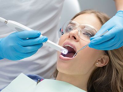
Intraoral cameras are compact, pen-sized devices designed to take high-resolution color images inside the mouth. Using a small lens and built-in lighting, these cameras provide a close-up view of teeth, gums, and other oral structures that is far more detailed than what the naked eye can see during a routine exam. Modern devices are lightweight, easy for clinicians to maneuver, and capable of delivering sharp still images as well as short video clips for review.
The captured image is displayed instantly on a chairside monitor, allowing both the clinician and the patient to examine the same view at the same time. This real-time visual feedback helps clinicians identify subtle surface changes, hairline fractures, early-stage decay, and soft-tissue concerns that might otherwise be missed. Many intraoral cameras also include macro focus and angled tips to reach difficult areas comfortably.
Because the camera records in full color and high definition, it provides reliable documentation that supports clinical decision-making. The level of detail a camera delivers makes it an excellent adjunct to visual inspection and radiography, complementing other diagnostic tools rather than replacing them. For dental teams, that means more confidence in diagnosing conditions and proposing appropriate care paths.
One of the clearest advantages of intraoral cameras is how they transform clinical findings into visual information patients can easily understand. Instead of relying on descriptions or diagrams alone, patients see precisely what the dentist sees—an exact image of a stained groove, a chipped edge, or inflamed gum tissue. That shared perspective reduces confusion and helps people make informed choices about their oral health.
When clinicians pause to review images with patients, it encourages questions and opens a collaborative conversation about treatment options, preventive strategies, and daily care. Visual evidence also helps set realistic expectations; patients can better appreciate the stages of disease, the rationale for recommended procedures, and the importance of follow-up. That clarity often leads to improved adherence to home care instructions and scheduled visits.
In addition, intraoral images are useful teaching tools during exams and consultations. Practitioners can point to specific features—such as enamel wear patterns or the margin of an existing restoration—and explain why those findings matter. For many patients, seeing a clear image is more motivating than hearing a general recommendation, because the need for care becomes tangible and immediate.
Intraoral cameras enhance diagnostic accuracy by revealing details that can be difficult to detect with visual inspection alone. Surface cracks, early carious lesions, and marginal breakdown around restorations are easier to identify with magnified, well-lit imagery. This level of detail supports earlier intervention, which can preserve more natural tooth structure and lead to simpler, less invasive treatments.
Beyond detection, these images are valuable when planning restorative and cosmetic procedures. Precise views of tooth anatomy and surrounding tissues help clinicians design restorations that fit better and look more natural. During complex treatment planning—such as full-mouth reconstruction or multi-unit restorative work—combining intraoral images with radiographs and digital impressions gives a more complete picture of oral health.
Intraoral cameras also contribute to continuity of care. Clear photographic records make it easier for a dental team to track changes over time, monitor healing after procedures, and assess the durability of restorations. When multiple clinicians are involved—whether within the practice or at a specialist referral—shared images support consistent treatment decisions and smoother coordination.
Captured intraoral images become part of the patient’s clinical record, stored securely within the practice’s imaging system. These images serve multiple purposes: documentation of baseline conditions, evidence of progressive change, and verification of completed treatments. Organized image archives allow clinicians to compare past and present findings quickly and to document the rationale behind clinical recommendations.
When appropriate and with the patient’s consent, these images can be shared with dental specialists, laboratories, and other members of the care team to facilitate cooperative treatment. A high-quality photograph can communicate nuances of tooth shape, shade, and tissue contours more effectively than a verbal description alone, improving the accuracy of lab-fabricated restorations and the efficiency of specialist consultations.
Privacy and data security are central when storing and transferring clinical images. Practices use secure systems designed to protect patient information and comply with applicable regulations. Patients who have questions about how their images are handled should feel comfortable asking the dental team for an explanation of the practice’s privacy and consent procedures.
Using an intraoral camera is a quick, noninvasive part of a dental exam. The clinician will guide the small camera into the mouth, directing it toward areas of interest while the patient remains seated comfortably. The device’s lighting and compact design minimize discomfort and make it easy to visualize hard-to-see surfaces, such as the back molars and the spaces between teeth.
Most imaging sessions last only a few minutes. The clinician may capture several still photos or brief video clips to illustrate different angles and findings. After the images are taken, the clinician will review them with the patient on the chairside monitor, explain any noteworthy observations, and discuss next steps. This review is an opportunity to ask questions and to clarify the recommended course of care.
Because intraoral imaging is supportive rather than definitive on its own, clinicians combine camera findings with other diagnostic tools—clinical assessment, probing, and radiographs—before making treatment decisions. Patients can expect the dental team to use the images as part of a larger assessment that aims to deliver accurate diagnoses and personalized, evidence-based care.
Intraoral cameras have become a standard part of modern dental practice by improving diagnosis, enhancing patient communication, and supporting thorough clinical documentation. At Clifton Modern Dentistry, we use contemporary imaging tools to help patients understand their oral health and to guide careful, well-informed treatment planning. If you’d like to learn more about how intraoral imaging is used in our office, please contact us for more information.


An intraoral camera is a compact, pen-sized imaging device that captures high-resolution color pictures and short video clips inside the mouth. It uses a small lens, built-in lighting, and often macro focus to produce detailed views of teeth, gums, and other oral structures. Clinicians maneuver the camera to target specific areas and record images that are displayed instantly on a chairside monitor.
The device provides magnified, well-lit views that are far more detailed than what the naked eye can see during a routine exam. These images serve as a visual adjunct to clinical inspection and radiography, helping to reveal subtle surface changes and areas of concern. Many modern intraoral cameras include angled tips that make it easier to reach back molars and tight spaces comfortably.
Intraoral cameras enhance diagnostic accuracy by revealing fine surface details that can be difficult to detect with direct vision alone. They make it easier to identify hairline cracks, early-stage decay, marginal breakdown around restorations, and soft-tissue abnormalities. The combination of magnification and consistent lighting reduces the likelihood of missed findings during an exam.
Because images are recorded in high definition and stored in the patient record, clinicians can compare findings over time and corroborate visual observations with radiographs and clinical probing. This layered approach supports earlier, less invasive interventions when appropriate and strengthens the evidence base for recommended treatments. Clear photographic documentation also improves consistency across multiple clinicians involved in care.
Yes. Intraoral imaging is noninvasive and does not use ionizing radiation; it relies on optical photography and internal lighting to capture images. The devices are designed to be small and lightweight, minimizing discomfort while allowing clinicians to visualize hard-to-see areas like the back molars and interproximal spaces. Most patients find the process quick and well tolerated, with individual imaging sessions typically lasting only a few minutes.
Dental teams follow infection control protocols by using disposable sleeves or barriers and cleaning the device between patients to maintain hygiene. Because the procedure is primarily observational, it is suitable for a wide range of patients, including children and seniors, and can often be performed as part of a routine exam without special preparation. Patients with strong gag reflexes or other concerns should discuss comfort strategies with their clinician beforehand.
Intraoral images provide detailed visual information that helps clinicians plan restorative and cosmetic procedures with greater precision. Photographs clarify tooth anatomy, the condition of existing restorations, and tissue contours, which aids decisions about margins, material selection, and shade matching. When combined with digital impressions and radiographs, these images contribute to a comprehensive clinical picture that improves the fit and esthetics of final restorations.
For complex treatment plans, clinicians use intraoral images to communicate specifics to dental laboratories and specialists, reducing ambiguity and improving outcomes. Clear photographs can show critical nuances—such as cusp form, contact relationships, and soft-tissue height—that guide laboratory work and chairside adjustments. This cooperative workflow supports predictable, evidence-based treatment planning and coordinated care with consultants when needed.
Intraoral cameras transform clinical findings into clear visual information that patients can easily understand, which improves informed decision-making. Instead of relying solely on verbal descriptions, patients see exact images of issues like staining, chips, or inflamed tissue, which helps clarify the nature and extent of a problem. Reviewing images together fosters a collaborative conversation about preventive strategies and treatment choices.
The visual evidence also sets realistic expectations by showing the patient the current condition and explaining why particular treatments are recommended. Clinicians can point to specific features in the image to illustrate causes and consequences, which often motivates better adherence to home care and scheduled follow-ups. Using images as a teaching tool makes oral health priorities tangible and actionable for patients.
An intraoral imaging session typically takes only a few minutes and is easily incorporated into a standard dental exam. The clinician will guide the small camera around the mouth to capture several still photos or brief video clips from different angles. Patients remain seated comfortably while the images appear immediately on the chairside monitor for joint review.
After capturing images, the clinician will review notable findings with the patient and explain recommended next steps, which may include monitoring, preventive measures, or further testing like radiographs. Because camera findings are used alongside other diagnostic tools, patients should expect a comprehensive evaluation that combines visual images with clinical assessment and radiographic information when appropriate. The review period is a good opportunity to ask questions and clarify the proposed plan.
Captured intraoral images become part of the patient’s clinical record and are stored in the practice’s secure imaging system alongside other documentation. These images provide baseline records, evidence of change over time, and visual confirmation of completed treatments. Organized digital archives allow clinicians to retrieve and compare past and present findings quickly when monitoring progress or evaluating outcomes.
Patient privacy and data security are important considerations when storing and transferring clinical images, so practices use secure systems and follow applicable privacy regulations. Images are shared with outside providers or laboratories only with patient consent and through secure channels to protect confidentiality. Patients who want details about how their images are handled should feel comfortable asking the dental team to explain consent and storage procedures.
No; intraoral cameras are a complementary diagnostic tool rather than a replacement for radiographs. Cameras capture detailed surface and soft-tissue images that are excellent for identifying visible cracks, staining, and restorative margins, but they do not show internal tooth structure, bone levels, or interproximal decay beneath the enamel. Radiographs remain essential for assessing bone health, underlying decay, and structures that are out of view of optical imaging.
Clinicians typically use intraoral images together with radiographs, clinical probing, and digital impressions to form a complete diagnostic picture. Each modality contributes different information, and the combined data set supports more accurate diagnoses and better-informed treatment plans. Patients can expect the dental team to explain how and why specific imaging methods are used for their particular needs.
Intraoral images help clinicians evaluate the subtle details that influence restorative and cosmetic outcomes, such as the margins of existing restorations, enamel wear patterns, and tissue contours. Photographs provide precise visual references for shade selection, shape, and surface texture, which improves communication with dental laboratories and guides chairside adjustments. Clear images reduce ambiguity when specifying the form and finish of crowns, veneers, and other restorations.
During treatment planning, images are used to document the initial condition and to guide procedures so that final results integrate naturally with adjacent teeth and soft tissues. They also aid clinicians in monitoring healing and evaluating the success of restorations over time, contributing to more predictable restorative and esthetic care. Accurate visual documentation supports better functional and cosmetic decisions for individual patients.
Patients can view intraoral images on the chairside monitor during their appointment, where the clinician will review findings and explain any recommended next steps in detail. With the patient’s permission, images can also be saved to the record for later review or printed or exported for sharing with a specialist or a dental laboratory. Reviewing these images together helps ensure patients understand their condition and the rationale behind any proposed care.
If you have questions or would like a more detailed consultation, contact Clifton Modern Dentistry at our office on 310 Terrace Ave in Cincinnati, OH, or call New Patients at (513) 214-3739 or Existing Patients at (513) 285-8055 to schedule an appointment. Our team can explain how intraoral imaging will be used in your exam and arrange a time for a chairside review with a clinician if you prefer a focused discussion of specific concerns.

Have Questions or Need an Appointment?
Getting in touch with Clifton Modern Dentistry is simple! Call us or use our online form, and our friendly team will help you schedule visits, answer your questions, and guide you toward the best care for your smile.
Don’t wait—start your journey to a healthier, brighter smile today!